Molecular Imaging Techniques:
X-ray Crystallography
Multi-dimensional NMR (Indirect)
Electron Microscopy (TEM / SEM)
We began with X-ray crystallography and 3D - NMR techniques. Here we will present another imaging tool - electron microscopy. There are several excellent web tutorials for electron microscopy and images to illustrate the power of the techniques. Our coverage of electron microscopy will be limited to TEM and SEM. Our discussion of imaging methods concludes with a brief introduction of four common medical imaging techniques: CAT / MRI / and PET scans and the use of ultrasound.
Refer to the web sites for further information.
I. General Principles of a Electron Microscope:
How does an EM work?
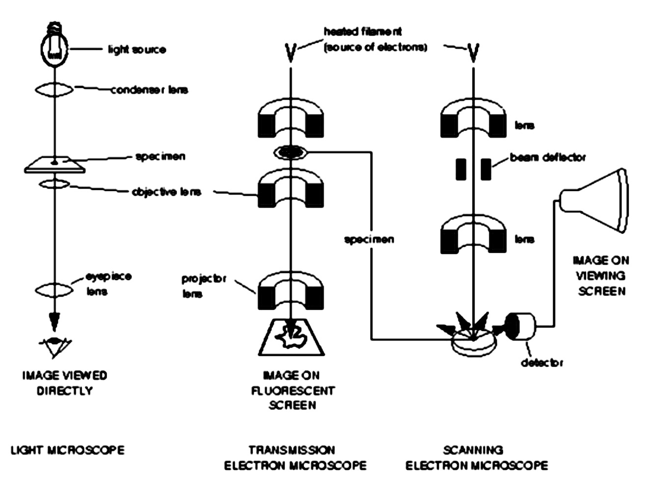
There are many different kinds of electron microscopes, but all use a beam of electrons as a "light" source and electromagnetic lens for focusing purposes. Our study will be limited to TEM (transmission electron microscopy) and SEM (scanning electron microscopy). TEM is very similar to light microscopy or a slide projector, SEM is quite different as noted below:
Figure above is after Fig. 1-6 from an introduction to EMs of many types found at: http://www.mih.unibas.ch/Booklet/Lecture/Chapter1/Chapter1.html
Here is a web site at Texas A&M that describes many frequently used EMs in their facility - http://www.tamu.edu/mic/instruments.html
II. TEM - Transmission Electron Microscopy
TEM requires fairly thin specimens to allow the electron beam to penetrate the sample. Much of the earlier work relied on stains (heavy metal compounds) to introduce contrast. In "negative" stain TEM procedures, a sample was deposited on a Cu grid coated with plastic in a thin film of heavy metal stain. Contrast could be seen between the path of the electron beam through regions of the stain that contained or did not contain the heavy metal.
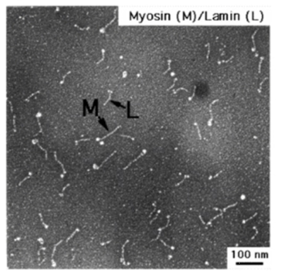
Today the images of many viruses and protein complexes are being reconstructed by collecting data on frozen samples taken at many orientations. Hundreds of these individual images are analyzed, grouped, averaged and studied using concepts of self-assembly and molecular symmetry and then "reconstructed" with the aid of a computer to generate 3D images of these large particles. Here are two links to image libraries of such reconstructed images:
Example: 3D image reconstruction of rhinovirus complexed with ICAM-1 receptor
For more images - try Professor Michael Rossmann's Laboratory Home Page or Prof. Tim Baker's Home Page.
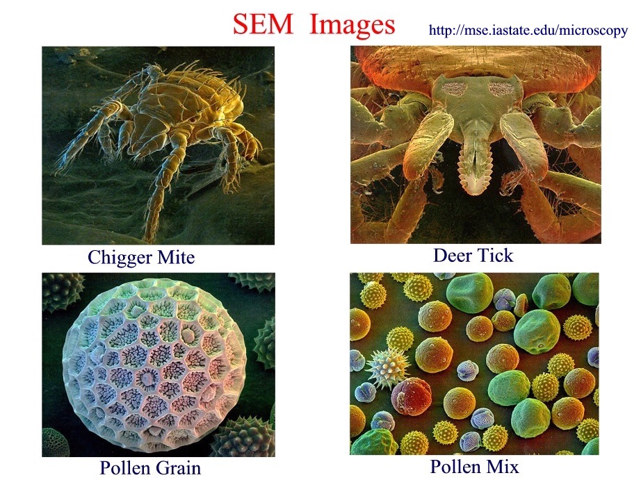
III. Scanning Electron Microscopy
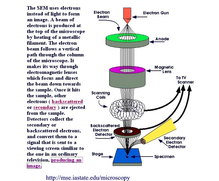
There are excellent tutorials on SEM aimed at the grade school, high school and college level at this web site from the Materials Science and Engineerign Department at Iowa State University:
http://mse.iastate.edu/microscopy/home.html
Shown below is a summary figure from the tutorial illustrating the essential components of a SEM.
The Iowa State web site also features a "Picture Library" of SEM images, here is but a sample of the kind of images that can be produces by SEM:
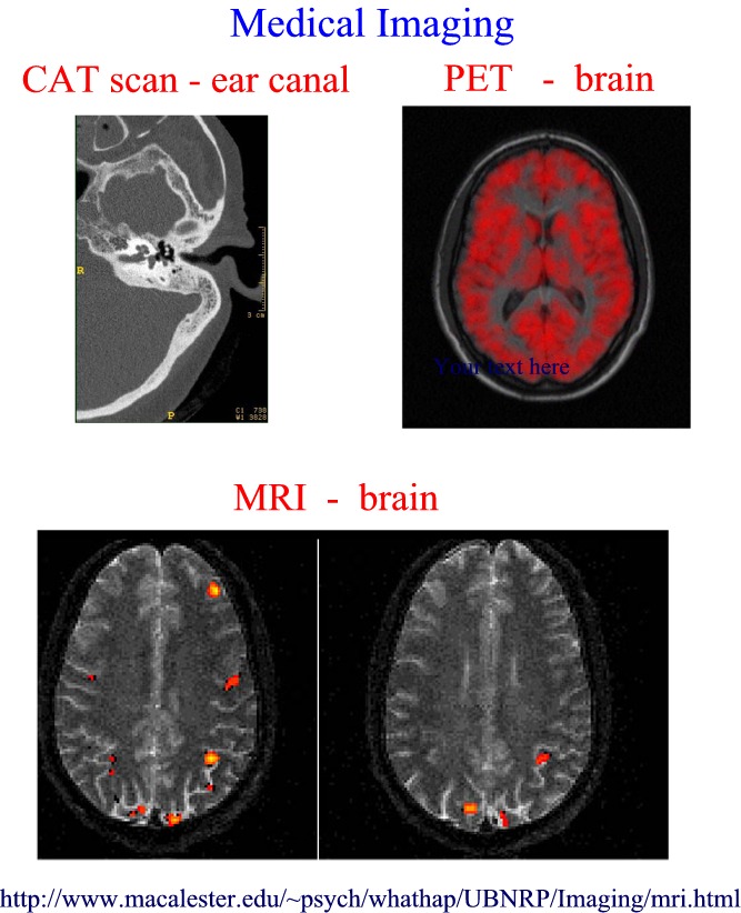
IV. Medical Applications
Imaging is also very important in medicine. Know the names and principles behind these common methods used in medical applications:
.Medical Imaging Techniques: Tutorial Images
CAT scans
- A CT scan is essentially a computerized assembly of several x-ray images taken from a series of different angles. With a CT, the resolution is much better than conventional x-rays, and the detail that can be seen is much greater. As with all other typical x-rays, the procedure is radiographic and the patient's body is exposed to a small
amount of radiation during the scan.
MRI - (Nuclear) Magnetic Resonance Imaging is a noninvasive imaging technique that does not use x-rays (unlike CAT scan). The process involves passing a strong magnetic field through the head. The magnetic field used is 30,000 + times that of the earth's magnetic field. It's effect on the body, however, is harmless and temporary. The MRI scanner can detect radiation from certain molecules, which are present in different concentrations in different tissues. The fluid contrast between structures in the brain can then be visualized.
PET
- Positron Emission Tomography, or PET, is a procedure that allows a physician to
examine the heart, brain, and other organs. PET images show the chemical functioning
of an organ or tissue, unlike X-ray, CT, or MRI which show only body structure.
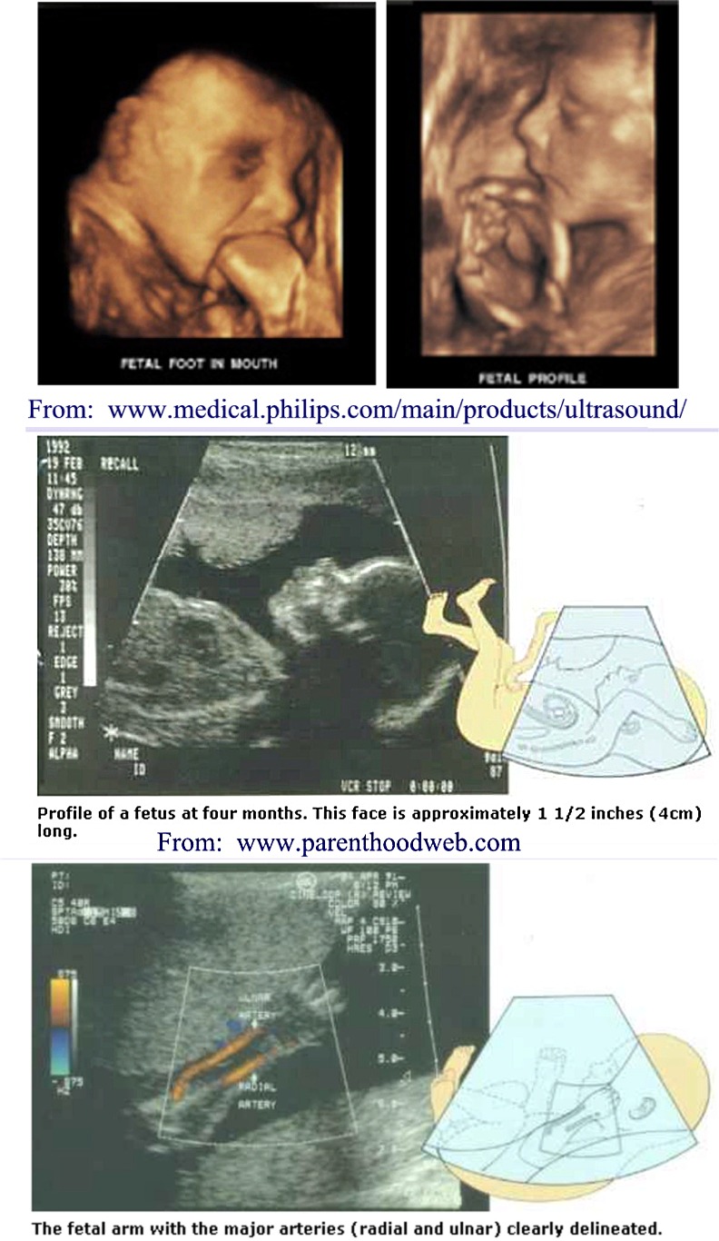
Sonography - The use of 3D - ultrasound as a procedure to image fetuses, and various tissues. Ultrasound scanners can be regarded as a form of 'medical' Sonar. "SONAR" refers to Sound Navigation and Ranging. A detailed history of the development of the use of ultrasound can be on the web at this site.
There are many web sites for learning more about medical imaging and radiology. Here is one that describes each of the above techniques (CT / MRI / PET) and provides images as examples, and another site with fetal ultrasound images.