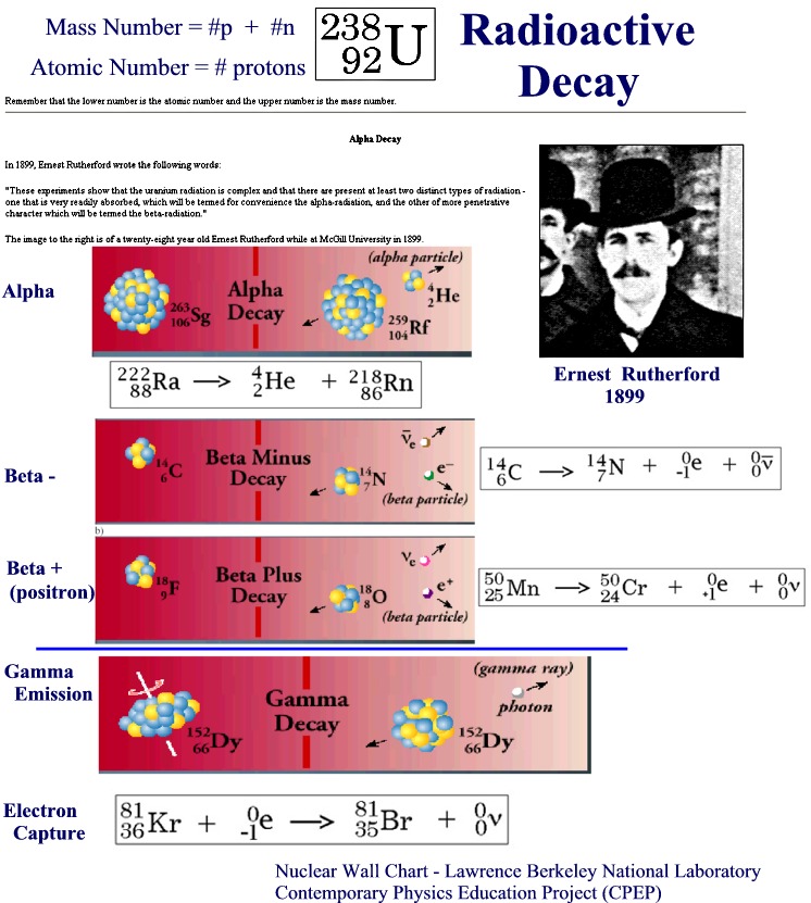
Nuclear decay by emission of an alpha particle (4He nucleus ).
Our coverage of radioactivity will be limited to the following six topics:
1. Review of the Nuclear Atom:
a) Chemistry Model:
Electron - An elementary particle with a unit electrical charge and a mass 1/1837 of the proton. Electrons surround the atom's positively charged nucleus and determine the atom's chemical properties.
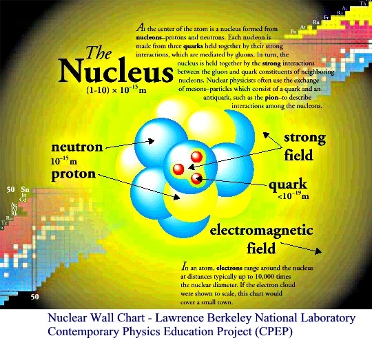
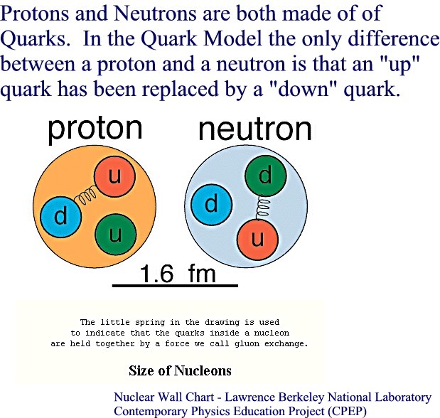
Proton - One of the basic particles which makes up an atom. The proton is found in the nucleus and has a positive electrical charge equivalent to the negative charge of an electron and a mass similar to that of a neutron. A proton is a hydrogen nucleus.
Neutron - One of the basic particles which make up an atom. A neutron and a proton have about the same weight, but the neutron has no electrical charge.
Neutrino - An electrically neutral particle with negligible mass. It is produced in many nuclear reactions such as in beta decay.
b) Nuclear Physics Model:
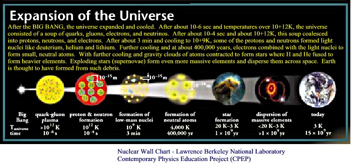
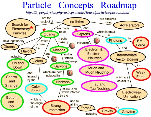
The results of particle accelerator experiments have led scientist to postulate the existence of three types of forces important in the nucleus: The strong force, the weak force, and the electromagnetic force. These forces are though to account for all types of interaction found in matter. The fourth force found in nature, but not in the nucleus, is the gravitational force. Quarks are particles with no internal structure that are thought to be the fundamental constituents of hadrons. Neutrons and protons, or nucleons, are hadrons that are thought to be composed of three quarks each. In the BIG BANG theory, the universe consisted of a soup of quarks, gluons, electrons and neutrinos for a fraction of a second before neutrons and protons formed. (Aside: "The Particle Zoo" and a Roadmap to Particle Concepts)
2. Review of Isotopes
Table of Isotopes (Berkeley Laboratory) - Isotopes: Two or more nuclides having the same atomic number, thus constituting the same element, but differing in the mass number. Isotopes of a given element have the same number of nuclear protons but differing numbers of neutrons. Naturally occurring chemical elements are usually mixtures of isotopes so that observed (non-integer) atomic weights are average values for the mixture.
Of the nuclei found on Earth, the vast majority is stable. This is so because almost all short-lived radioactive nuclei have decayed during the history of the Earth. There are approximately 270 stable isotopes and 50 naturally occurring
radioisotopes (radioactive isotopes). Thousands of other radioisotopes have been made in the laboratory. Light nuclei are most stable when the ratio of neutrons/protons ~ 1 (C-12). Heavier nuclei are most stable when the neutron to proton ratio is >1 (~1.5); e.g. U-235 (Z=92, ratio = 1.55) --> Pb 208 (Z=82, ratio = 1.54)
3. Types of Radioactive Decay:
Some history in the development of the study of radioactivity:
1895 that x-rays were discovered and Radiology was born.
1896, Becquerel amazed to find that photographic plates had been subjected to intense radiation produced by Uranium salts.
1898 Marie Slodovska Curie and her husband Pierre discovered Polonium and Radium and called the new form of energy, that was emitted, as Radioactivity. (See "Women in Chemistry" in the Spring 2000 "Chemical Compositions" newsletter, p.4)
1899 Ernest Rutherford discovered alpha and beta forms of emitted Radiation.
1928 - Geiger-Muller Counter - perfected.
1970 - Freedman proposed and built a rotating tomographic γ-camera. Radiologists developed the word tomography from the Greek words «tome» meaning to cut and «graphy» meaning to write.
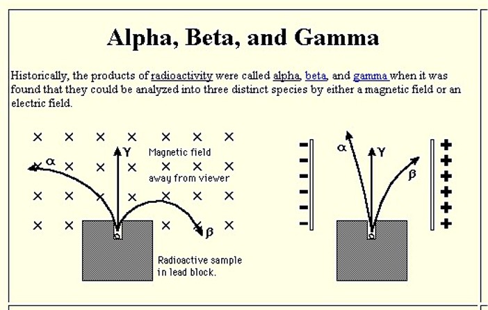
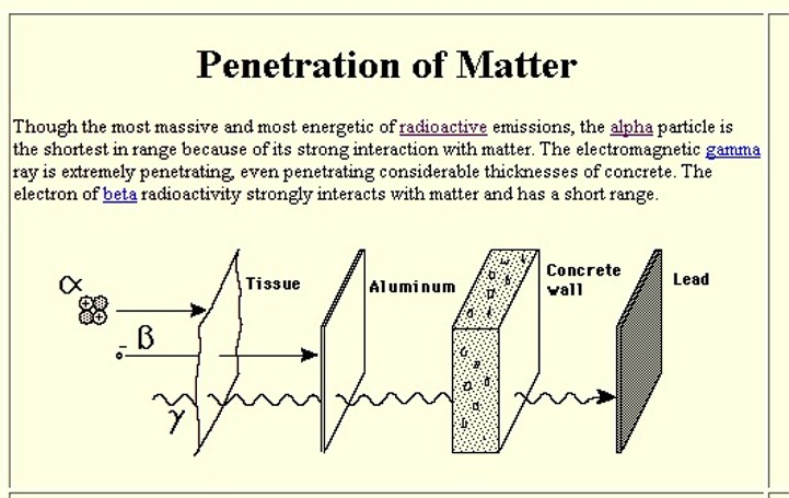
Common types of radioactivity - these differ in their charge and penetrating / ionizing power.
Alpha
Decay![]() (click
here for animation)
(click
here for animation)
Nuclear decay by emission of an alpha particle (4He nucleus ).
Beta
Decay![]() (click
here for animation)
(click
here for animation)
Nuclear decay by emission of an electron or a positron. Positron decay is always
accompanied by electron capture decay. (Aside: What is a Neutrino?)
Gamma
Rays![]()
A highly penetrating type of nuclear radiation, similar to x-rays and light,
except that it comes from within the nucleus of an atom, and, in general, has a
shorter wavelength. Gamma rays emission is a decay mode by which excited state
of a nucleus de-excite to lower (more stable) state in the same nucleus.
Electron
Capture Decay![]()
Nuclear decay by capture of an atomic electron. If the decay energy is
greater than 1022 keV, positron emission can also occur in competition with
electron capture.
4. Review of Half-life equations / C14 dating
Half-Life - Used to measure the rate of radioactive decay of disintegration. The time lapse during which a radioactive mass loses one half of its radioactivity.
A = Ao exp(-kt) where k = ln2/half-life
C-14 (5730 years); I-131 (8.02 days); Tc-99m (6.01 hours)
5. Ways to Count Radioactivity:
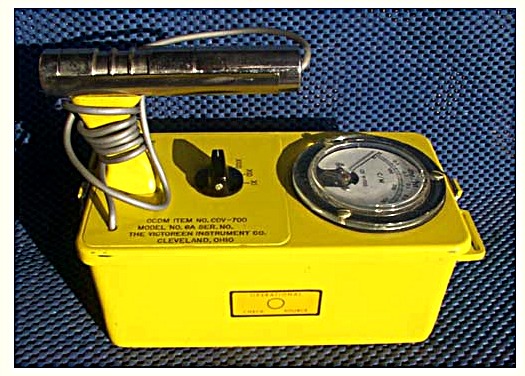
Film - The first recording of radiation was made using film. It is still used many places today. It took about 30 years before the Geiger Counter was developed.
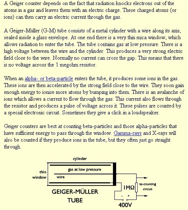
Geiger Counter - A radiation detector consisting of two electrodes with a low-pressure gas (argon or neon) in between. A high voltage is maintained such that if radiation passes through the counter, it ionizes the gas resulting in an avalanche of electrons generating the signal. Geiger counters count radiation but cannot distinguish either the energy or kind of radiation.
Scintillation Counter
A scintillation counter consists of a material that emits light when radiation
passes through it. Various liquid, plastic, and crystalline materials have
scintillation properties. Scintillation light is measured with photomultiplier
tubes. In general the amount of scintillator light detected is proportional to
the energy of the radiation. Liquid scintillation counting where the
radioactive sample is mixed with a scintillation "cocktail" is a
common biochemistry technique. (More notes
on ionization detectors)
Semiconductor Detector
Radiation striking very pure Ge and Si semiconductor detectors can excite a
large number of electrons into the conduction band leading to a measurable
current. This current is proportional to the energy of the radiation.
Semiconductor detectors can be used to accurately measure the energy and
intensity of radiation.
Autoradiography with the PhosphorImager
Radiation striking a storage phosphor screen can store latent images from
radioisotopes that can later be "read" using a PhosphorImager system.
This system has a scanning helium-neon laser (red light) that is focused to a spot at the plane of the phosphor screen. The beam sweeps across the screen. As the screen is scanned, the BaFBR:EU+2 crystals in the screen release
energy as blue light phosphorescence and return to the ground state. The
blue light emitted is collected by a fiber optic bundle and
channeled to a photo multiplier tube to form a quantitative
representation of the sample. These phosphorimager screens have the added
advantage that they are more sensitive
than film and can be "cleared" for reuse by simple exposure to bright,
white light.
5. Applications / Terms: Use of Radioactivity in Science and Medicine
Units of Radioactivity and Dose
Original unit - amount of radioactivity was the
curie (Ci) - activity of one gram of radium-226.Today 1 curie = 3.7 10+10 radioactive decays per second [exactly].
International System of Units (SI) the becquerel (Bq) has replaced the curie, where
1 becquerel = 1 radioactive decay per second = 2.703 10
-11 Ci.
The magnitude of radiation exposures is specified in terms of the
radiation dose.There are two important categories of dose:
1. The
absorbed dose, sometimes also known as the physical dose, defined by the amount of energy deposited in a unit mass in human tissue or other media. The original unit is the rad [100 erg/g]; it is now being widely replaced by the SI unit, the gray (Gy) [1 J/kg], where 1 gray = 100 rad.2. The biological dose, sometimes also known as the dose equivalent, expressed in units of rem or, in the SI system, sievert (Sv). This dose reflects the fact that the biological damage caused by a particle depends not only on the total energy deposited but also on the rate of energy loss per unit distance traversed by the particle (or "linear energy transfer"). For example, alpha particles do much more damage per unit energy deposited than do electrons. This effect can be represented, in rough overall terms, by a quality factor, Q. Over a wide range of incident energies, Q = 1.0 for electrons (and for x-rays and gamma rays, both of which produce electrons) and Q = 20 for alpha particles. For neutrons, the adopted quality factor varies from 5 to 20, depending on neutron energy. The biological impact is specified by the dose equivalent H, which is the product of the absorbed dose D and the quality factor Q: dose equiv. H = Q D.
The unit for the dose equivalent is the rem if the absorbed dose is in rads and the sievert (Sv) if the absorbed dose is in grays. Thus, 1 Sv = 100 rem. 1 rem is roughly the average dose received in 3 years of exposure to natural radiation. 1 Sv is at the bottom of the range of doses that, if received over a short period of time, are likely to cause noticeable symptoms of radiation sickness.
(Related notes on Radioisotopes)
**********************************************
Radiation Imaging is also very important in medicine. Here are three methods commonly involved medical applications:
.Medical Imaging Techniques (more on this later in the semester):
CAT scans
- A CT scan is essentially a computerized assembly of several x-ray images taken from a series of different angles. With a CT, the resolution is much better than conventional x-rays, and the detail that can be seen is much greater. As with all other typical x-rays, the procedure is radiographic and the patient's body is exposed to a small
amount of radiation during the scan.
MRI - (Nuclear) Magnetic Resonance Imaging is a noninvasive imaging technique that does not use x-rays (unlike CAT scan). The process involves passing a strong magnetic field through the head. The magnetic field used is 30,000 + times that of the earth's magnetic field. It's effect on the body, however, is harmless and temporary. The MRI scanner can detect radiation from certain molecules, which are present in different concentrations in different tissues. The fluid contrast between structures in the brain can then be visualized.
PET - Positron Emission Tomography, or PET, is a procedure that allows a physician to examine the heart, brain, and other organs. PET images show the chemical functioning of an organ or tissue, unlike X-ray, CT, or MRI which show only body structure.
There are many web sites for learning more about medical imaging and radiology - send me the best ones you find and I will add them to our class links..